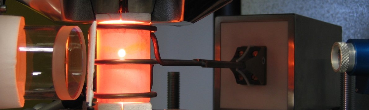
Research Areas

Functionalization and Assembly of Ceramic Nanoparticles

Bioceramics

Novel antibacterial and abrasion resistant ceramic microstructures

Biofouling and erosion of material surfaces are two of the major challenges in fluid transport or food processing systems which contain micro-abrasive particles. Biofouling represents a serious public health risk as uncontrolled growth of pathogenic microorganisms such as bacteria may cause serious epidemic outbreaks and mass diseases. Micro abrasion of material surfaces caused by particles in the fluid system diminishes the service life of fluid handling devices. The aim of this research project is the fabrication of novel biofunctionalized microstructured surfaces that kill bacteria and prevent biofilm formation on the surface by combining different antibacterial agents. The material surfaces shall be microstructured in order to increase the abrasion resistance, resulting at the same time in the protection of the immobilized biomolecules on the surface. The application of such engineered surfaces addresses aqueous systems with possible usage in fluid transport devices and food processing chains where biofouling must be prevented.

Microstructure

Micro-structured biofunctionalized ceramic surfaces and their impact on cells

The main objective of this research project is to understand cell interactions with ceramic materials as a function of surface microstructure and surface functionalisation. Different micropatterning techniques and biomolecule modifications are applied to elucidate cell adhesion, proliferation, migration and viability on calcium phosphate surfaces. The findings of these fundamental investigations are aimed to improve the bioactivity of medical implants such as ceramic-coated artificial joints, dental implants and ceramic bone scaffolds. Examples of different microstructure techniques are given in the figures left.

Cells

Cell evaluation of novel microporous Hydroxyapatite-based scaffolds for bone replacement

The development of materials that are suitable to replace hard or soft tissue is a field of intense research. However, there is still no material to date that features all the properties of natural bone at the same time: on the one hand the excellent mechanical properties, on the other hand a composition and optimal microstructure suitable for the three-dimensional ingrowth of bone cells. The ideal bone replacement material must exhibit a similar fracture strength and fracture toughness to bone. At the same time, however, it should be bioresorbable in order to be replaced by bone tissue in the long term. Hydroxyapatite resembles bone apatite to a high degree, but is not as soluble. We use hydroxyapatite as the main component for our ceramics, and change the composition in order to enhance its properties towards an ideal bone replacement material. Bone cells do not only react towards the chemical composition of the material itself, but amongst others to surface structure, micro- and macroporosity and adsorbed or bound proteins, respectively peptides. At the present, we examine growth of osteoblasts and biochemical reactions as a function of chemical composition, surface roughness and microporosity. After having found the optimal conditions, macropores will be introduced to enable three-dimensional growth of the cells and subsequently the mechanical properties of the resulting material will be evaluated.

scaffolds

Inorganic/organic scaffold composites for bone tissue engineering with gradual porosities

The human bone structure is complex and includes on the one hand dense structures referred to as Compacta and on the other hand sponge-like structures called Spongiosa. The open porosity of bone represents an additional challenge. At the same time, the specific chemical composition of bone, consisting of a mineral phase (bone apatite) and an organic phase (collagen), provide its biological functionality. This research project focuses on both aspects, the morphological and the chemical/biological adaption of bone properties. On the long term, the processed scaffolds need to feature bioactivity and controlled biodegradation with a sufficient initial mechanical stability. To fabricate a Compacta-like structure we use Freeze Cast Processing (in collaboration with the Ceramics group), providing pores of 10 to 20 microns. To adapt the structure of the Spongiosa, we apply a Polymer Replica Method. By this method, pore sizes can be adjusted from 100 to 1000 microns. In a next step, the obtained structures are biofunctionalized and cell tested in order to assess the bioactivity. The mechanical and degradation properties are elucidated before, during and after cell tests.

Nanodiamonds 

Nanodiamonds

Nanodiamonds are a fascinating material that only recently came into the spotlight despite being known for over sixty years. Recent breakthroughs in the last decade regarding processing, purifying, and dispersing nanodiamonds enabled widespread research on diamond nanoparticles and quickly showed the inherent potential of this newly rediscovered nanomaterial. Among the outstanding properties of the sp3 hybridized carbon nanoparticles are inherent fluorescence, excellent biocompatibility and ease of surface functionalization. Moreover, pure detonation nanodiamonds are easy to procure without being prohibitively expensive. Based on these features, nanodiamonds are starting to be intensely investigated as promising candidates for biomedical applications like drug delivery, nanoparticle-assisted diagnostics and imaging, or as implant coatings and reinforcements. Nanodiamonds have generally been considered biocompatible for a broad variety of eukaryotic cells.
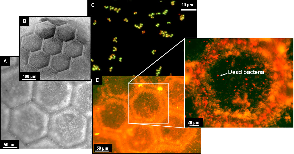
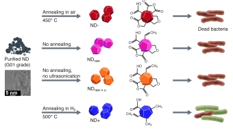
In recent work, we showed that, depending on their surface composition, nanodiamonds kill Gram-positive and -negative bacteria rapidly and efficiently. We investigated six different types of nanodiamonds exhibiting diverse oxygen-containing surface groups that were created using standard pretreatment methods for forming nanodiamond dispersions. Our experiments suggest that the antibacterial activity of nanodiamond is linked to the presence of partially oxidized and negatively charged surfaces, specifically those containing acid anhydride groups. Furthermore, proteins were found to control the bactericidal properties of nanodiamonds by covering these surface groups, which explains the previously reported biocompatibility of nanodiamonds. We are currently starting to collaborate with several institutes to further elucidate the antibacterial surface properties of nanodiamonds. These collaborations will enable the detailed analysis of the surface chemistry of antibacterial nanodiamonds using XPS, Raman and NMR, as well as computational methods that model the surface/biomolecule interactions of nanodiamond particles. In another 
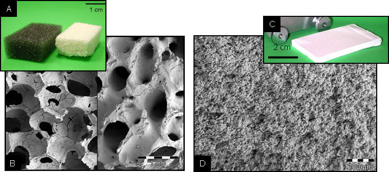
project, we are incorporating nanodiamonds into bone replacement materials based on hydroxyapatite in order to provide these biomaterials with antibacterial properties.


Colloidosomes

Colloidosomes are hollow capsules formed via self-assembly of colloidal particles on emulsion droplets. Consequently, they consist merely of a cohesive shell of nanoparticles. This type of semipermeable capsule possesses great potential for the encapsulation and sustained release of active agents, such as drugs, flavors, or fragrances. We developed a straightforward method which for the first time allowed the preparation of submicrometer-sized colloidosomes with tailorable nanopores. Capsule formation is carried out at mild pH, ambient temperature, and without the use of hazardous chemicals. In combination with a lipid that carries the same net charge at an oil−water interface as the colloidosome-forming particles in aqueous media, we are able to synthesize colloidosomes with positive and negative zeta potentials. The capsules are stable both in organic as well as in aqueous environments. By varying the sizes and shapes of the nanoparticles, we were able to tailor the pore diameters and pore size distributions on the surface of the capsules. Tailoring the size of the colloidosome nanopores potentially allows the controlled release of encapsulated active agents of different size, such as proteins, antibiotics or chemotherapeutics. Since basically any kind of nanoparticles can be used for the fabrication of colloidosomes, they present an extremely versatile platform for multifunctional microcapsules.

Currently, we are working on the preparation of submicron colloidosomes that incorporate different kinds of functional nanoparticles. Recently, we achieved the combined assembly of colloidosomes from superparamagnetic iron oxide nanoparticles and fluorescent silica particles (Figure 3). Furthermore, using nanodiamonds, colloidosomes with high shell stability could be formed that represent a new platform for the preparation of more complex, multifunctional capsules which we called diamondosomes.

Carbonate CarbonateCarbonate 

Biomimetic Formation of Calcium Carbonate Microcarriers and Micropatterned Ceramics

Based on a novel approach that takes into account the coacervation of calcium and poly(acrylic acid) (PAA), we are able to biomimetically produce bulk quantities of amorphous calcium carbonate (ACC) particles (Figure 4). Coacervation is a phase separation mechanism which is caused by the complexation of lyophilic, polyionic polymers with counterions and results in the formation of a colloidal droplet phase. We study the time- and concentration-dependent growth of Ca2+/PAA coacervate droplets using dynamic light scattering (DLS), turbidity and titration measurements. Applying these results to the generation of high amounts of unstable ACC particles, we are able to produce highly concentrated ACC suspensions. This processing route represents a versatile platform for the bottom-up preparation of micropatterned ceramics on the basis of calcium carbonate.

The same approach can be utilized to prepare calcium carbonate microcarrier particles by choosing appropriate salt concentrations and reaction parameters. Currently, we are studying the release of active agents from these mineralized coacervates . Furthermore, we are working on transferring the coacervation method to other minerals to produce functional biomimetic materials. So far, we have been successful with preparing calcium phosphate and magnetite/maghemite spheres (d ≈ 200 nm). Our next target are zinc oxide particles. In general, this method provides a mild way (moderate pH, room temperatre) to prepare large amounts of homogeneous and monodisperse inorganic nano and micro-particles with good control over particle size and composition.


Mineralized Collagen Nanofibers

The fundamental approach of this project is the preparation of mineralized fibers via the membrane method . Due to the strong supersaturation and close proximity of both calcium and phosphate ions as well as collagen molecules at the exits of the pores of a track etched membrane, amorphous calcium phosphate nucleates in the constrained interstitial spaces between collagen molecules. The resulting fibrils show the characteristic banding pattern that is a result of the staggered array of collagen molecules within the fibril and closely resemble mineralized collagen fibrils found in mammalian bone. The figure shows examples of mineralized fibrils that were prepared using the membrane method. Note that the banding pattern of the fibrils would not be visible using TEM without contrast agents; instead, here, calcium phosphate provides sufficient contrast. This procedure is currently the only known bottom-up method for the preparation of mineralized collagen. The membrane method allows precise control of the diameter of the fibrils by choosing the pore size of the membrane. We investigate methods to mineralized collagen fibrils with alternative materials like superparamagnetic iron oxide nanoparticles or silver nanoparticles to introduce additional functionality to these fundamental building blocks of bone.


Rheology of Hydroxyapatite Suspensions

The synthesis of porous hydroxyapatite scaffolds is essential for biomedical applications such as bone tissue engineering and replacement. One way to induce macroporosity into hydroxyapatite scaffolds, which is needed to support bone in-growth, is to use protein additives as foaming agents. Another reason to use protein additives is the potential to introduce a specific biofunctionality to the synthesized scaffolds. In this project, we studied the rheological properties of a hydroxyapatite suspension system with additions of the proteins bovine serum albumin (BSA), lysozyme (LSZ) and fibrinogen (FIB). Both the rheology of the bulk phase as well as the interfacial shear rheology were studied. The rheological properties of interfacial films can be studied by either dilatational or interfacial shear rheology. Though dilatational rheology of droplet systems is often used as a model for foam behavior, it does not provide structural information on the interfacial film, since Gibbs and Marangoni effects play a huge role for the elasticity of the interface. The advantage of interfacial shear rheology is the ability to measure the time dependent growth of interfacial films in situ while gaining access to the structural information that is regularly revealed by oscillatory rheological methods.